Glaucoma- Be Aware of the Dangers
Share this Image On Your Site
Embed This Infographic on Your Site:
Glaucoma represents a group of diseases of the eye that cause injury to the optic nerve and a subsequent loss of vision. While there are several types, the three most common are open-angle glaucoma, closed-angle glaucoma, and normal-tension glaucoma. The disease can present gradually with few symptoms, or suddenly with many. Initially, peripheral vision is decreased, after which there is a failure of central vision and total blindness.
There are multiple risk factors for glaucoma, including having hypertension, obesity, migraine headaches, increase pressure within the eye itself, and a positive family history of glaucoma. Some people will have undiagnosed high blood pressure (of at least 21 mm Hg) without actually knowing about it until their vision is affected. Others will have optic nerve injury suggestive of glaucoma but won’t have high pressures inside the eye. The main cause of glaucoma is a decreased rate of exit of the eye’s aqueous humor through the trabecular meshwork (drainage channels) in the anterior chamber of the eye. An eye examination with special tests can identify the high pressure and optic nerve damage.
Glaucoma is not curable but it is possible to decrease the progression of the disease by various means, such as laser treatment, eye drops, oral drugs, and rarely, surgery. Each of these treatments can be used to decrease the internal pressure of the eye. Most people only need eye drops; however, some patients will get better using laser eye surgery or one of several glaucoma surgeries, especially if medications fail. If glaucoma develops suddenly, emergency measures must be taken to decrease the progression of vision loss.
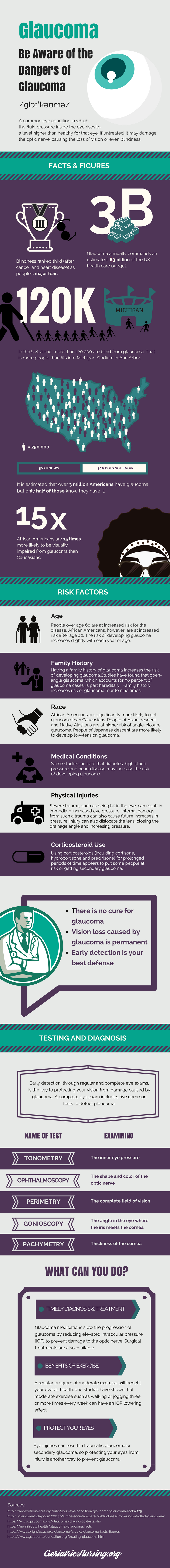
In the US, about 3 million people have glaucoma with only half of all patients having the disease knowingly. About 120,000 individuals are considered blind from the disease, making up about 10 percent of all cases of complete blindness. The problem is more common in developing countries so that glaucoma represents the second most common cause of blindness throughout the world. There is a greater incidence of the disease among people of African descent, causing the second most common kind of blindness in this population, fifteen times more likely to occur in this group.
Open angle glaucoma represents the most common kind of glaucoma, causing 19 percent of cases of blindness in people of African descent and 6 percent of cases in Caucasians. People who especially need glaucoma screening are very nearsighted people, diabetics, individuals over 60 years of age, and those with a known family history of glaucoma. It is believed that more than 60 million people are at risk for glaucoma around the world.
Types of Glaucoma
Glaucoma come in several types, the most common of which are identified by having an increase in intraocular pressure (IOP), which is the actual pressure inside the aqueous portion of the eye. More than 90 percent of all glaucoma cases can be identified as open-angle glaucoma. The cause of this type of glaucoma stems from clogging of the trabecular meshwork, the drainage canal of the eye, resulting in a buildup of fluid and increased eye pressure.
The examination shows an “open angle” at which the cornea meets the iris. This angle has a certain width in normal people and increased pressure develops gradually over time with few symptoms in the beginning. It is also referred to as chronic glaucoma (as it is a chronic, lifelong disease) or primary glaucoma. It is believed to affect three million people in the US, only some of whom know they have the disorder.
Angle-closure glaucoma is the second most common type. The incidence of this type of glaucoma is about 4.2 out of 100,000 people per year in the total population and about 11 out of 100,000 people per year in those over the age of 30 years. It is caused by an acute blockage of the trabecular meshwork (drainage canals) of the eye, resulting in a sudden increase in intraocular pressure. There is a narrow angle between the cornea and iris. The symptoms are usually quite noticeable and progress very rapidly. Emergency ophthalmic care is necessary to control the high eye pressure and prevent permanent vision loss. Some people refer to it as narrow-angle glaucoma or acute glaucoma.
Normal-tension glaucoma or NTG is also called normal-pressure glaucoma or low-tension glaucoma. In this type of glaucoma, there is the typical damage to the optic nerve without any findings of high blood pressure in the eye itself. No one knows the exact reason why glaucoma changes happen in this disorder in the face of normal intraocular eye pressure. Vision loss is similar to the more common causes of glaucoma.
Low-tension glaucoma cannot be diagnosed by assessing the intraocular eye pressure but is instead found through direct observation of the optic nerve using an ophthalmoscope. A visual field test can also be done that looks for peripheral nerve deficits typical of glaucoma. Patients with early disease don’t always recognize the peripheral vision defect. It is estimated that up to 25 percent of patients with primary open-angle glaucoma (POAG) experience low-tension glaucoma.
Primary congenital glaucoma happens to babies when there is an abnormality or incomplete development of the drainage system (trabecular network) of the eye in utero. This is a relatively rare eye condition that might be hereditary. The incidence of primary congenital glaucoma (PCG) is about 1 in 10,000–18,000 live births. The incidence depends strongly on the patient’s ethnicity and whether or not they were born from a consanguineous union. Surgery can be done to correct the problem, helping to restore vision.
Secondary glaucoma can be open-angle glaucoma or angle-closure glaucoma and can occur in just one or both eyes. It is referred to as “secondary” because it stems from some other injury or illness in the eye. The treatment sometimes depends on taking care of the primary cause of the eye problem and sometimes on treating the high blood pressure inside the eye.
Exfoliative glaucoma happens when there is flaky material that peels off the outer part of the lens inside the eye. This flaky material gathers in the anterior chamber of the eye and clogs the trabecular meshwork, resulting in a decreased drainage and increased pressure in the eye. Some refer to it as pseudoexfoliative glaucoma. This type of glaucoma is more prevalent among certain ethnic groups, including those who have ethnicity stemming from Russia, the Nordic countries, Greeks, Mediterranean populations and East Indians. A genetic defect has recently been associated with this particular condition, but it does not develop in childhood or at birth.
Neovascular glaucoma is caused by the unwanted accumulation of new blood vessels (neovascularity) in both the iris and the trabecular network of the eye. It is never found in isolation but is linked to diabetes and other diseases. The neovascularization results in a blockage of the fluid from the trabecular meshwork, resulting in high pressure inside the eye.
Pigmentary glaucoma happens when the granules of pigment behind the iris of the eye break off into the aqueous humor of the eye, flow towards the trabecular network, and ultimately block the network, clogging the drainage canal resulting in an increased eye pressure. Research has revealed that vigorous exercise can result in more pigment released from the iris, which can worsen the degree of eye drainage. Patients with pigment dispersion syndrome or pigmentary glaucoma should reconsider performing heavy exercise unless their doctor tells them it is okay to do it.
Traumatic glaucoma usually affects just one eye and is the result of a direct injury to the eye. This is a type of secondary open-angle glaucoma that can occur suddenly and immediately following the eye injury or may develop many years later. It is generally secondary to blunt trauma to the eye but can be from some type of penetrating injury.
Uveitic glaucoma stems from having uveitis, an inflammation and swelling of one of the middle layers of the eye, called the uvea. Its normal job is to supply blood to the retina. When the uvea becomes inflamed, it blocks the trabecular meshwork. Uveitis can cause increased intraocular pressure when inflammatory debris inhibits the flow of fluid through the trabecular meshwork. Over the long term, the inflammation itself can result in scar tissue in the eye that further obstructs fluid outflow; treatment with corticosteroids can also result in increased intraocular pressure.
Irido-corneal endothelial syndrome can result in secondary glaucoma. Also called ICE, this disease stems from cells on the back side of the cornea spreading over the trabecular network and across the surface of the iris. This results in an increase in intraocular pressure, which can damage the optic nerve. The spread of these cells also causes adhesions (scar tissue) to form, which cause adherence of the iris to the cornea, further blocking the trabecular network.
Risk Factors & What to Look for
Because the signs and symptoms of glaucoma can be subtle or absent, it is important to have an awareness of the risk factors for the condition. Anyone who is older than sixty years of age is automatically at risk for the disease. People with known high intraocular pressure will have a greater risk for developing glaucoma. Those individuals with a family history of glaucoma will be at a greater risk for the disorder as will anyone from African-American or Hispanic origin. Being extremely nearsighted will predispose a person to the disease as will certain health problems, such as sickle cell anemia, hypertension, coronary artery disease and diabetes. Eye injuries and past eye surgery will increase the risk of the disease. Women with estrogen deficiency early in life will have a greater chance of developping glaucoma. Corticosteroid eye drops will increase the risk as well.
Causes of Glaucoma
The ultimate cause of glaucoma is an imbalance between the amount of aqueous humor (eye fluid) in the eye and the amount of fluid that drains through the trabecular network out of the eye. If this imbalance occurs, the intraocular (inner eye) pressure will increase, damaging the optic nerve and affecting vision. The exact reasons behind the blockage of the trabecular network differ according to the type of glaucoma a person has.
Aqueous humor is made behind the iris, begins flowing through the anterior chamber through the pupil of the eye, and leaving the eye through the drainage angle via the trabecular network located between the iris and cornea. If too much aqueous humor is being made or too little flows through the trabecular network, the amount of fluid builds up, increasing intraocular pressure (IOP). This pressure can increase dramatically and dangerously to ultimately result in optic nerve damage and glaucoma. The fluid actually presses on the optic nerve, damaging the delicate nerve fibers. Because of the way the optic nerve fibers are arranged, peripheral vision is lost first, followed by central vision.
As previously mentioned, glaucoma can be open-angle or closed-angle (narrow-angle). The narrower the angle in the anterior chamber, the harder it is for the aqueous humor to flow through the trabecular network. Patients can also have open-angle glaucoma if there is something, like debris or cells blocking the outflow tract of the aqueous humor. Scar tissue can also adversely affect the outflow of fluid from the eye.
Some people can have glaucoma associated with normal pressure, also referred to as normal-tension glaucoma. These people seem to have optic nerve fibers particularly sensitive to pressure and they will suffer from irreversible damage in the face of what is found to be normal intraocular pressure. Interestingly, some patients will have increased intraocular pressure (a problem referred to as ocular hypertension) but will not have glaucoma and won’t ever develop it as a complication. Because IOP is not always associated with glaucoma, the optic nerve needs to be evaluated and the visual field testing to make sure there aren’t glaucoma complications in the face of normal intraocular pressure.
It is believed that normal-tension glaucoma is somehow related to decreased blood flow to the optic nerve. There can be vasospasm or other causes of decreased blood flow to the optic nerve, increasing the sensitivity of the nerve fibers, resulting in symptoms associated with glaucoma and permanent optic nerve damage. Amyloid deposits seen in Alzheimer’s dementia also build up in the retina, which may sensitize the optic nerve to pressure damage.
Diagnosis of Glaucoma
The best way to diagnose glaucoma is to have a regular eye exam that includes a measurement of the intraocular pressure and an optic nerve inspection. Ideally, a person should have this type of examination every 2-4 years prior to the age of 40 years, every 1-3 years between the ages of 40 years and 54 years, every 1-2 years between the ages of 55 and 64 years, and every 6-12 months after the age of 65 years. High risk individuals need to be examined for glaucoma approximately every year after the age of 35 years.
There are five basic tests for glaucoma any one of which might not make the diagnosis alone without a complementary test. The intraocular pressure is measured with tonometry; the features of the optic nerve are evaluated using ophthalmoscopy (with dilation of the pupils); perimetry or visual field testing can assess the patient for a visual field defect; gonioscopy measures the angle between the cornea and iris; the thickness of the cornea is measured with pachymetry testing.
Tonometry is one of the two main tests for glaucoma and measures the intraocular pressure (the pressure inside the eye). During tonometry testing, the surface of the eye is first anesthetized with a numbing eyedrop. A device called a tonometer is placed on the surface of the eye or a small puff of air is blown onto the eye surface with the pressure recorded. A normal intraocular pressure is between 12 and 22 mm Hg or “millimeters of mercury”. Glaucoma can be suspected in any person who has an intraocular pressure of at least 20 mm Hg. Remember, too, that normal-pressure glaucoma can exist so that other testing is necessary to determine whether the disease exists, particularly if the patient has symptoms.
The second most common test used to identify glaucoma is the ophthalmoscopy examination. This is a diagnostic test that helps the eye doctor look carefully at the optic nerve, looking specifically for damage to the optic nerve fibers. Eye drops are first given to cause dilation of the pupil so the entire retina can be seen with the ophthalmoscope. The device is shined onto the eye and the doctor looks at the optic nerve to see if it is normal. If there is an abnormality in the appearance of the optic nerve or an identifiable ocular hypertension, the patient should have further evaluation with additional testing.
Perimetry testing is a maps the patient’s visual fields and looks specifically for peripheral field defects seen in early glaucoma. The patient looks straight ahead during the test and follows a moving light beam that passes through their peripheral field of vision. Defects are mapped on paper to see if there is peripheral loss of vision or, in more severe cases, blindness in the central part of the visual field. Visual field testing is often done repeatedly throughout the course of diagnosed glaucoma as it is a good measurement of the progression of the disease. It can be done every 6-12 months in documented glaucoma cases to check the patient’s visual fields.
Gonioscopy is not done to screen for glaucoma but is used to tell the difference between narrow-angle glaucoma and wide-angle glaucoma. The treatment of each is slightly different and the causes of each are different. The test is done by first anesthetizing the surface of the eyes and placing a contact lens on the cornea. The lens contains a mirror on the back of it that can be used to highlight the angle between the cornea and the iris. This angle can be measured to identify the type of glaucoma the patient has.
The test referred to as pachymetry is a painless examination that evaluates the thickness of the cornea, which is the central clear membrane in front of the eye. A pachymeter or probe is placed on the cornea and is used to measure the cornea’s thickness. It can help diagnose glaucoma and dictate treatment because the treatment and intraocular pressure readings can depend on the cornea thickness. Thicker corneas are less flexible and it will be more difficult for the eye to adjust to high pressures.
Modern pachymeter devices make use of ultrasound technology, while earlier devices were once based on optical principles. Ultrasonic pachymeters have traditionally been devices that measure the total thickness of the human cornea in the form of a number in micrometers displayed on the readout. The newest generation of ultrasonic pachymeters work using a technology called Corneal Waveform (CWF). This technology allows the ophthalmologist to obtain an ultra-high definition “echogram” of the cornea and its thickness.
Because it is not always a simple task to identify glaucoma, more than one test is generally necessary to make the diagnosis. Tonometry can pick up many cases of primary glaucoma; however, there is a subset of glaucoma cases that have normal intraocular pressures, so that ophthalmoscopy and peripheral field testing needs to be done to evaluate the patient at high risk for glaucoma who has normal tonometry readings.
Signs and Symptoms of Glaucoma
While some patients will have no symptoms, there are early symptoms that can identify a patient who has or who may develop glaucoma at some point in their life. The symptoms of angle-closure (narrow-angle) glaucoma and open-angle glaucoma are different. Patients with open-angle glaucoma don’t often have any identifiable symptoms in the beginning of the disease process, so examination of the patient’s eyes is especially important to detect glaucoma before blindness develops.
The main symptoms of open-angle glaucoma include a gradual vision loss, particularly in both eyes, and mainly involving peripheral vision first. As the disease progresses untreated, the patient eventually develops tunnel vision and complete blindness.
Patients with acute angle-closure glaucoma have a sudden onset of increased intraocular pressure, resulting in an ophthalmological emergency necessitating urgent treatment to avoid blindness, which can develop within one to two days after the onset of symptoms. The typical signs and symptoms of angle-closure glaucoma are more dramatic than open-angle glaucoma and include severe pain in the affected eye, secondary nausea and vomiting, and the acute onset of decreased vision that often starts with difficulty seeing things in low light conditions. Vision can be blurry and there will be halos seen around lights. On examination, the affected eye will have conjunctival injection, appearing to be red in all parts of the sclera.
The symptoms of chronic angle-closure glaucoma are not as dramatic as acute angle-closure glaucoma, with a gradual reduction in vision starting in the periphery of the visual field and optic nerve damage that progresses to tunnel vision with symptoms nearly identical to open-angle glaucoma. People with normal-tension glaucoma will generally not experience any eye pain or other visual symptoms with the exception of an early loss of peripheral vision that progresses over time.
Prevention
There is no cure for glaucoma, so the best treatment is to prevent eye damage early in the course of the disease process. This means that even the asymptomatic patient should be evaluated for glaucoma prior to the age of 40 years by having ophthalmological examinations including tonometry testing every 2-4 years. After the age of 40 years and up to the age of 54 years, exams should take place every 1-3 years. From the age of 55 to the age of 64, eye exams should take place every 1-2 years. Because the incidence of glaucoma increases with age, anyone over the age of 65 should have an examination of the eyes every 6-12 months. Patients with a family history of glaucoma or known ocular hypertension should be examined every 1-2 years, starting at the age of 35 years. People with siblings who have glaucoma, diabetics, and people of African descent are considered high risk and should be examined earlier in life.
There is actually no way to prevent the onset of glaucoma, but things like peripheral vision loss and blindness can be minimized by recognizing and treating the disease as early as possible. Primary open-angle glaucoma has a relatively slow disease progression and few symptoms in the early stages so that if ocular hypertension is identified by tonometry, stepped-up surveillance can begin that will allow for early treatment before peripheral visual changes develop or progress to a loss of central vision. Medications can be given as soon as the disease is identified (or surgery can be performed) so that the disease never progresses past the early stages.
Researchers have uncovered the fact that performing regular, moderate aerobic exercise will not only benefit the patient’s overall health status but will decrease the intraocular pressure. The downside is that the benefits of this form of exercise only persist as long as the patient is actively exercising. For this reason, it is recommended that the patient jog or walk at least three types per week. There has been an additional benefit for patients who perform yoga, although doing inverted poses will increase the intraocular pressure, resulting in the worsening of glaucoma.
Eye protection will prevent trauma to the eye that might ultimately result in traumatic glaucoma. Any type of eye injury, including blunt trauma and penetrating eye trauma, can damage the eye and affect the trabecular meshwork’s ability to drain aqueous humor from the eye, damaging the optic nerve and causing secondary glaucoma.
While the research on foods to prevent glaucoma is slim, there is some evidence that certain foods will decrease the risk of glaucoma. Dark green, yellow, and orange fruits and vegetables contain carotenoids, which may protect the eyes from developing several eye disorders, including glaucoma. Lutein and zeaxanthin are particularly important for the health of the eyes. These substances can be found in dark, leafy greens such as spinach, collard greens and kale, as well as in yellow corn, okra, Brussels sprouts, mango, sweet potatoes, lima beans, green beans, squash, green, yellow and orange bell peppers, broccoli, and egg yolks.
Treatment Options
There is a wide variety of treatments for glaucoma, including eye drops or pills that act to decrease the intraocular pressure in the eyes. The most common glaucoma treatment involves the use of eyedrops that are effective in treating glaucoma by decreasing the amount of aqueous humor produced by the eye in the first place or by increasing the outward flow of the fluid through the trabecular network. Often, more than one eyedrop is required to manage the disease.
There are several types of eyedrops for glaucoma. One of these is the alpha-adrenergic agonist drug, which reduces the production of aqueous humor and increases the outflow of the fluid through the trabecular network channels. The major downside of using this drug is that there is a high incidence of allergies with this classification of glaucoma medications. Typical examples of alpha-adrenergic drugs for glaucoma include apraclonidine, dipivefrin, epinephrine and brimonidine.
Beta-blocker eyedrops can also be used to manage glaucoma. This classification of drugs works by decreasing the production of aqueous humor and decreasing the influx of fluid into the eye itself, thereby reducing intraocular pressure. Commonly-used drugs of this class include betatoxol, metipranolol, carteolol, levobunolol and timolol.
Carbonic anhydrase inhibitors are another classification of drugs used to manage glaucoma. They can be eyedrops or oral medications that decrease the fluid production within the eye, decreasing the intraocular pressure. The different drugs of this class include dorzolamide and brinzolamide (which are eyedrop medications), and acetazolamide or methazolamide (oral glaucoma drugs).
Miotic drugs for glaucoma are cholinergic agonists, which cause constriction of the pupil. This results in an increase in the fluid drainage out of the eye. Some examples of miotic drugs for glaucoma include pilocarpine and echothiopate drops.
Prostaglandin analog medications will decrease the intraocular pressure by increasing the outflow of aqueous humor through the trabecular meshwork. Examples of this classification of drugs include tafluprost eyedrops, latanoprost drops, bimatoprost, travoprost and unoprostone ophthalmic solution.
Because there is often the need for more than one type of eyedrop type in the treatment of glaucoma, a number of combination eyedrops can be used to decrease the number of eyedrops. These include combination drops consisting of dorzolamide and timolol, brinzolamide and brimonidine, brimonidine and timolol, and latanoprost and timolol.
Nowadays, the most common non-drug treatment for glaucoma involves performing one of several different laser procedures designed to decrease intraocular pressure. The downside of these treatments is that the decreased pressure in the eye does not last a lifetime, with increases in intraocular pressure occurring later on in certain patients, depending on their age at the time of treatment, the kind of glaucoma the patient has, and any other predisposing medical conditions. Many patients will still need medical therapy but won’t need as much medication to affect a decrease in IOP.
Laser therapy can be used in patients with angle-closure glaucoma, open-angle glaucoma, or neovascular glaucoma. The eye is numbed and the laser is directed at different parts of the eye. When used on the trabecular meshwork, it can increase the rate of outflow of the aqueous humor. The laser can also be directed at the retina, to benefit the optic nerve, the ciliary body or the iris itself.
A laser procedure called an argon laser trabeculoplasty or ALT is used to treat patients who have open-angle glaucoma. In ALT, the laser is directed specifically at the trabecular meshwork. Clogged canals within the meshwork are opened up so that fluid can pass through the canals and out of the eye, decreasing intraocular pressure. Another procedure called selective laser trabeculoplasty or SLT uses a lower energy laser targeting certain cells of the trabecular meshwork, accomplishing essentially the same thing as an ALT procedure but without collateral damage to healthy tissue.
A laser peripheral iridotomy procedure or LPI is used to treat angle-closure glaucoma. In this disease, the angle between the cornea and iris is too narrow blocking the fluid from exiting the eye. This procedure uses a laser to make a hole in the iris to allow fluid to flow out of the eye through a different pathway than the trabecular meshwork.
Patients with aggressive or extremely severe open-angle glaucoma can be effectively treated with cyclophotocoagulation, especially when other treatments fail. The procedure involves using a laser directed endoscopically (or directly through the sclera) at the ciliary body, the part of the eye that makes the aqueous humor. This will decrease the eye pressure (at least temporarily) by decreasing the amount of fluid created by this structure.
Scatter panretinal photocoagulation is another glaucoma laser procedure. It acts to decrease the excessive vascularity that damages the optic nerve in patients who have neovascular glaucoma. Like all laser eye surgery, it can result in temporary blurry vision, secondary cataract formation, and irritation of the eyes.
Surgery is less commonly used for the treatment of glaucoma as compared to laser surgery. These conventional surgical techniques are generally reserved for instances when medical therapy and laser surgery have been unsuccessful in having sustained reductions in intraocular pressure. The choice of surgical treatment depends on how severe the disease is, the responsiveness to other treatment strategies, and any underlying health problems.
The main surgical procedure used to treat glaucoma is the trabeculectomy. This involves making an incision in the sclera of the eye, removing a section of the trabecular meshwork near the base of the cornea, and closing the incision. The effect is to surgically create a larger opening through which the aqueous humor can finally exit the eye, reducing intraocular pressure. One eye is treated at a time, with improvements so significant that many people no longer need to use eyedrops. The downside of the procedure is that the IOP can become too low or there will be scar tissue development that closes off the meshwork, necessitating a repeat surgery. Cataracts can be a late side effect of having a trabeculectomy.
In some cases, particularly in those who don’t do well after a simple trabeculectomy, the patient will respond to having a glaucoma drainage implant placed within the eye. In this procedure, a small tube is inserted directly in the anterior chamber of the eye between the lens and the cornea, maximizing the drainage of the aqueous humor of the eye.
A specialty surgical procedure for glaucoma uses a device known as the Trabectome™. It is used to treat patients with open-angle glaucoma by anesthetizing the eye, making a small incision in the cornea, and removing a section of the trabecular meshwork, which automatically increases the drainage in the eye. This is a low-risk procedure with few side effects.
Angle-closure glaucoma is often treated surgically using a peripheral iridectomy procedure. A small part of the peripheral portion of the iris is incised, allowing for drainage of aqueous humor through the excision site. There are also novel procedures used to decrease the IOP by increasing the outflow of fluid in the eye, such as canaloplasty and techniques that place different shunts and filtration devices into the drainage system of the eye.
Infants born with congenital glaucoma can be surgically treated with a goniotomy procedure, which is reserved just for this type of glaucoma. It involves using a small blade and making an incision in the cornea in order to cut through the trabecular meshwork. This increases the outflow of fluid from the eye and decreases intraocular pressure. These infants may also benefit from having a traditional trabeculotomy performed to cut through a blocked trabecular meshwork.
Additional Resources
What Is Glaucoma? This site is put out by the American Academy of Ophthalmology, making it an authoritative resource for a thorough discussion of the phenomenon of glaucoma, including the causes and symptoms of the disorder, the ways in which glaucoma is diagnosed and the treatment of this common disease of the eyes.
Facts about Glaucoma. The National Eye Institute, one of the nation’s top authorities on eye disorders puts out this website listing the major facts and questions the average person might have about glaucoma. Simple and complex questions on glaucoma are answered in easy layman’s terms along with easy-to-understand pictures on how the disorder starts, common symptoms of the disease, the way its diagnosed, and the available treatments.
Eyeing the 6 Early Warning Signs of Glaucoma.This website specifically looks into the early signs of glaucoma for people concerned they might have the eye disease and want specifics about what to look out for in terms of early symptoms. Six of the most common early signs of glaucoma are listed, including risk factors and ways the eye doctor can tell a person is at a higher than normal risk.
The American Optometric Association puts out this comprehensive website that includes the definition of glaucoma, the different types of the disease, the major signs and symptoms, diagnosis, and both the short-term and long-term treatment necessary to control the problem.
Juvenile Glaucoma. This is the right website for people who have or know someone who has juvenile glaucoma. The site talks about the different types of glaucoma seen in infants and children. The pathophysiology and etiology of this rare eye disease is discussed along with the workup and diagnosis of juvenile glaucoma and its treatment. This is a rare type of glaucoma and the site offers information not available from ordinary glaucoma websites.
What can I do to prevent Glaucoma? This web page discusses the important topic of glaucoma prevention. Most sites talk about the treatment of the disease but this is one of the few sites, put out by the Glaucoma Research Foundation, that actually talks about ways a person can prevent the development of glaucoma in the first place.
Types of Glaucoma. While it’s relatively easy to find a website that discusses the two main types of glaucoma, it’s harder to find one that covers all the major types of the disease. This webpage, put out by the Glaucoma Research Foundation, talks about the main types of glaucoma, including those most people haven’t heard of.
Foods to help reduce glaucoma risk. Few people realize that glaucoma risk can be reduced by eating the right foods. This article from Fox News provides astute insight into what is known about preventing glaucoma through eating right. It offers a great deal of insight into natural, dietary ways to keep at bay this common eye condition.
Medlineplus. This is the landing page for a number of comprehensive and diverse articles/webpages on the topic of glaucoma. The site offers glaucoma information in Spanish and talks about things like marijuana and the treatment of glaucoma, as well as glaucoma in minority groups.